
 Tiếng Việt
 Home
Home Introduction
 Announcements
 Student Essential
 Event attendance lists
Iron Deficiency Anemia (IDA) is the most common form of anemia worldwide, accounting for approximately 50% of all anemia cases. This condition is particularly prevalent among women of reproductive age, children, and the elderly.
A study conducted in the United States (2023) noted that the prevalence of iron deficiency among females aged 12–21 has not significantly changed over the past two decades, indicating that this remains a public health issue that needs to be addressed.
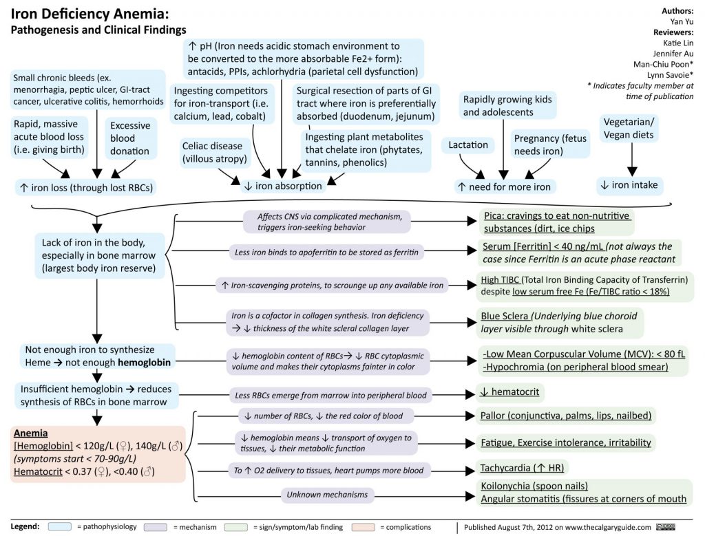
Iron deficiency anemia – pathology and specific manifestations
1. Pathophysiology
Iron is an essential component for hemoglobin (Hb) synthesis. When iron stores are depleted, erythropoiesis is affected through 3 stages:
- Stage 1 – Depleted stores: Ferritin decreases, without clinical manifestations.
- Stage 2 – Latent iron deficiency: Serum Fe decreases, TIBC increases, without anemia.
- Stage 3 – Iron deficiency anemia: Hb significantly decreases, microcytic hypochromic red blood cells.
Common causes include: chronic blood loss (menstruation, gastrointestinal bleeding), increased demand (pregnancy, rapidly growing children), and malabsorption (gastrectomy). Hepcidin – the hormone regulating iron metabolism – plays a central role in the pathogenesis by inhibiting iron absorption and its release from storage sites.
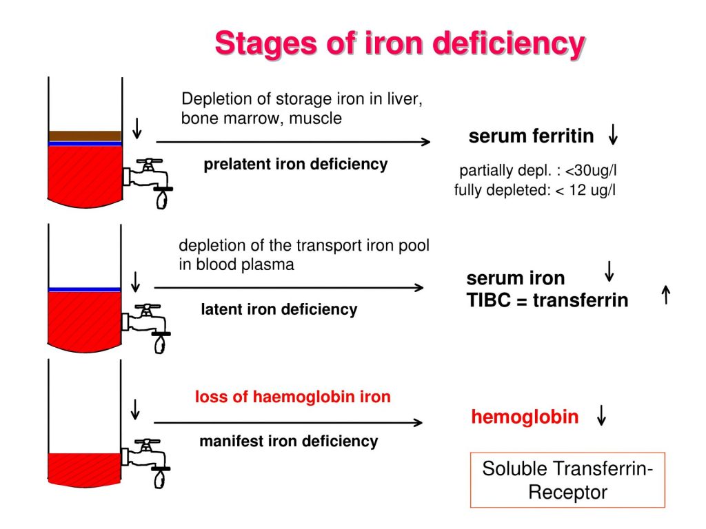
Stages of iron deficiency anemia
2. Clinical and Laboratory Findings
2.1. Clinical Symptoms
Patients often experience fatigue, pale skin, pale mucous membranes, palpitations, and dyspnea on exertion. Characteristic signs include: brittle flat nails (koilonychia), smooth red tongue, and difficulty swallowing (Plummer-Vinson syndrome). Neurological symptoms such as reduced concentration and irritability are also increasingly reported in recent medical literature [2].
2.2. Diagnostic Laboratory Tests
| Test | Male | Female |
| Hemoglobin | < 13 g/dL | < 12 g/dL |
| Ferritin | < 30 ng/mL | < 30 ng/mL |
| Serum Fe | < 55 µg/dL | < 40 µg/dL |
| TIBC | > 360 µg/dL | > 360 µg/dL |
| MCV | < 80 fL | < 80 fL |
Laboratory parameters in IDA by gender according to BSG 2021, AGA 2024, and WHO guidelines
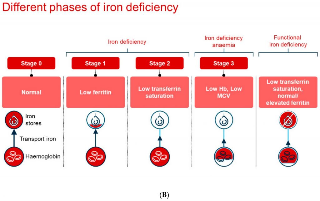
Laboratory findings in iron deficiency anemia
3. Treatment Approach
3.1. Etiological Treatment
The most crucial step is to identify and address the cause of iron loss. All male patients and postmenopausal women with IDA should undergo bidirectional gastrointestinal endoscopy to rule out gastrointestinal malignancies.
3.2. Oral Iron Supplementation (Preferred)
Ferrous sulfate is the preferred preparation due to its lowest cost. Current guidelines recommend once-daily dosing (instead of 2–3 times as previously), as studies on hepcidin show that lower doses optimize absorption. Additionally, vitamin C helps increase iron absorption. Duration of treatment: 3–6 months after Hb normalizes to replenish storage.
3.3. Intravenous Iron
Indicated in cases of: intolerance to oral administration, malabsorption, or a need for rapid recovery. Commonly used preparations include: Iron sucrose, Ferric carboxymaltose, and Ferric derisomaltose.
3.4. Blood Transfusion
Indicated when Hb < 7 g/dL or the patient presents with severe cardiovascular symptoms. It is not a routine treatment for uncomplicated IDA alone.
4. Monitoring and Key Takeaways
Check complete blood count after 4–8 weeks: an increase in Hb ≥ 1–2 g/dL confirms treatment response. Target ferritin after treatment should reach > 50 ng/mL. If there is no response, the diagnosis, treatment compliance, and underlying causes must be re-evaluated.
Key points to remember:
⁃ Low ferritin is the earliest and most sensitive marker of iron deficiency.
⁃ Always identify the root cause; do not just supplement iron while ignoring the underlying disease.
⁃ IDA in adult males → immediately suspect gastrointestinal bleeding.
⁃ Oral iron dosage is currently recommended once daily to optimize absorption via the hepcidin mechanism.
Dinh Toan Thang – Department of Medical Laboratory Technology